Last updated: Mon, Jul 8, 2024
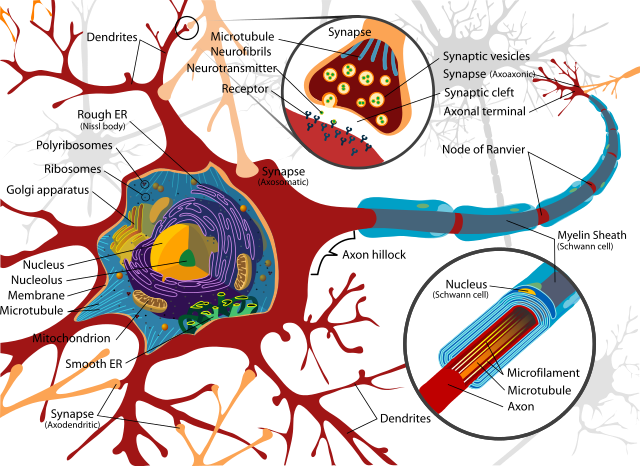
Pain is ultimately down to nerve cells or neurons. There is a wide variety of neurons in the body serving different functions, but we can use Figure 1: A neuron1 as an illustration (and an example of an excellent technical drawing). Neurons are cells that specialize in generating and sending signals. The bodies of neurons are highly variable in size. They range from 4 to 100 micrometers (millionths of a meter) in diameter. Based on 25 micrometers as a size, about 130,000 could be laid out in a single layer on a square inch, and 20,000,000 cell bodies would fit into a cubic inch.
Like most of the cells in your body, a nerve cell has a nucleus that contains chromosomes. Each chromosome contains genes (patterns used in manufacturing proteins), other patterns that are used to control the creation of proteins, and a lot more material whose function is not yet known. Each cell in an individual that contains a nucleus contains the same chromosomes and the same set of genes. A person's chromosomes contain, at recent estimates, about 20-25,000 genes.
Cells have different shapes and behaviors for two basic reasons: 1) Different genes are active in different cells, and 2) the cells have a different history. A gene that is currently being used to create proteins for the cell is said to be “active” or “expressed.” The history of a cell, then, is the history of which genes have been active at different points in the cell's lifetime.
The nucleus is in the main body of the cell, which is called the cell's soma. Close to the nucleus are specialized structures that build the proteins and other chemicals needed by the cell, and structures that break chemicals down either to harvest energy or to recycle chemical building blocks.
The nerve cell, like other cells, is separated from its neighbors and the fluids which bathe them by its cell membrane. The membrane is mostly made of a double sheet of lipids (fats). Because the membrane is fatty, it resists penetration by chemicals and compounds that dissolve readily in water. Some fats and proteins, on the other hand, can diffuse freely through the membrane. All the material within the membrane is referred to as cytoplasm.
A cell must sense and interact with its environment in order to maintain itself, and the cells that make up an organism must interact to coordinate their actions. Cells have a range of gadgets that, when installed in the membrane, they can use to selectively allow or force the movement of chemicals through the membrane. For example, cells normally use glucose as their source of energy. Glucose is distributed throughout the body by the circulating blood. Glucose won't penetrate the cell membrane by itself. The cell therefore makes glucose transporters that carry glucose molecules through the membrane. These transporters are proteins that are built using templates from the chromosome, and they are installed by the cell in its own membrane.
An ion channel is a protein that, when installed in the cell membrane, allows ions to move into or out of the cell. An ion is an atom or molecule that carries an electrical charge, either positive or negative. Sodium and chloride ions are the result when sodium chloride (table salt) is dissolved in water. Sodium ions carry a positive charge, while chloride ions carry a negative charge.
Cells must be able to control their ion concentrations to keep their machinery working correctly. Ions are especially important to neurons because of the electrical charge they carry. The electrical charge of ions is used by neurons to create their signals, as will be explained. If an ion channel were open all the time, ion concentrations inside the cell would become equal to concentrations outside the cell. For that reason, ion channels are either “gated” (meaning they can be open or closed) or “rectified” (meaning that ions can only pass in one direction). Ion channels are also selective to a specific type of ion. For example, an ion channel can be a sodium channel, or a potassium channel, and so forth. Cells are also equipped with ion pumps, which are protein machines that use energy from inside the cell to pump ions into or out of the cell. Using this variety of gadgets, the neuron can control the concentration of the various ions inside itself.
Another type of gadget that the cell uses to communicate with its environment is the receptor molecule. This is another type of protein machine that the cell builds and inserts in its own membrane. The portion of the receptor that is outside of the cell is shaped in such a way that it matches the shape of a specific molecule or class of molecules that may occur outside the membrane. The metaphor of lock-and-key is often used. The molecule that fits the receptor is called a transmitter if it is used by the body primarily to carry signals. (A more generic name for a molecule that fits a receptor is ligand, which means “something that binds.”) When a ligand binds to a receptor, it either causes the receptor to do something, or changes the way the receptor is functioning. For example, a particular transmitter may open an ion channel when it binds to it. Activation of other receptors can have major effects on the cell:
The body's hormones are transmitter molecules that bind with receptors on the various types of cells. Transmitters and receptors are also very important in the functioning of the nervous system. Transmitters used by the nervous system are called neurotransmitters.
Axons and dendrites are long protrusions or extensions of the cell body. They are enclosed by the same membrane that encloses the soma. They contain living cytoplasm, but are too small to contain the cell's protein machinery. Proteins and energy are distributed into the axons and dendrites either by diffusion or using a scaffolding of tiny tubules that works something like a tiny conveyor system. A neuron sends signals in a preferred direction. The axon is the extension that usually carries signals away from the soma. It is usually the longest of the extensions, too, although there is much variation in the shape of neurons. The dendrite or dendrites are usually heavily branched (“dendrite” is from the Greek word for tree), are usually shorter than the axon, and usually carry signals towards the soma. Axons and dendrites are both also referred to as “nerve fibers.” The branches at the ends of axons and dendrites are also confusingly called “fibers.” The final branches are called “terminal branches.”
The terminal branches of a neuron perform one of two functions. They either form synapses with another neuron, or they are sensory fibers that respond to stimulation such as pressure, temperature, or chemicals. A synapse is an area where two neurons come close together and can pass signals. Synapses can occur at the ends of terminal branches, along the axon or dendrite, or at the soma. This allows neurons to form complex networks and to influence each other in complex and sophisticated ways. Some of this will be elaborated on in subsequent sections.
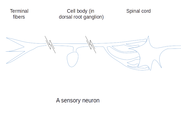
Figure 2 is a schematic view of a sensory nerve. In a sensory nerve, the axon splits into a “tee” shape after leaving the cell body. At one end of the axon are sensory (or terminal) fibers that respond to sensory stimuli. The sensory fibers may end in special sensory organs, or may be plain. At the other end are fibers that form synapses with neurons in the spinal cord or the brain stem.
The process by which a neuron emits or repeats a signal is called an action potential. The name refers to the electrical nature of the signal (an electrical potential is measured in volts). Although an action potential does have an electrical aspect, it isn't the same as an electric current running through a wire. An action potential uses the movement of ions to propel itself along the neuron's dendrites and axon, while a wire uses the movement of electrons. For that reason, an action potential moves much more slowly than a signal through a wire. Nevertheless, the action potential generates voltages that can be read by an electrocardiogram (which reads heart rhythms), an electromyelogram (which reads action potentials moving through muscle), or an electroencephalogram (which reads brain waves, action potentials moving through the cerebral cortex).
Neurons have ions in their own cytoplasm and are bathed in the extracellular fluid that bathes all the body's cells and is also mostly water. The ions that are most important in action potentials are small ions that move easily and quickly in the intracellular and extracellular fluids and that carry a large electrical charge for their weight. These ions include sodium, chloride, potassium, and calcium.
Ions can pass freely through the cell membrane only via ion channels or ion pumps. This allows the cell to control how many ions it contains. Where an ion moves, so moves its electrical charge, so the neuron can control its internal voltage relative to the surrounding fluid. A neuron at rest maintains its interior at a voltage that is about 90 millivolts (.09 volts) lower than its exterior, that is, -90 millivolts. It does this by pumping positive ions out. An action potential begins when the voltage of the neuron suddenly rises by about 25 millivolts. This normally happens at a synapse or at the sensors of a sensory cell. Once this threshold is reached, the ion channels open and allow positive ions to flood into the cell.
This action potential, assuming it is strong enough, tends to spread along the cell membrane in all directions to the limits of the cell. However, ion channels are distributed unevenly on the membrane in a way that encourages the action potential to travel toward the soma from dendrites or sensory fibers, and then away from the soma along the axon. As the action potential passes, the ion channels close and the ion pumps continue to pump. This restores the voltage to about -90 millivolts, and the cell is ready to generate another action potential. A single action potential can be over in about 0.2 milliseconds or .0002 seconds. Some neurons can repeatedly generate action potentials at a rate of over 1,000 per second, that is, a frequency of over 1,000Hz.
Sensory endings initiate action potentials in a variety of ways. Pressure-sensitive nerve endings, for example, contain channel-like membrane proteins that admit ions when they are distorted by pressure.
Action potentials can be transmitted from one neuron to another through the synapses between neurons. A synapse has an upstream neuron, called the presynaptic neuron, and a downstream neuron, the postsynaptic neuron. The signal normally goes from the presynaptic to the postsynaptic neuron. The synapse itself is made up of a presynaptic terminal and a postsynaptic terminal separated by a tiny gap called the synaptic cleft. (See Figure 1: A neuron.) When an action potential arrives at the presynaptic terminal, the presynaptic terminal releases a quantity of neurotransmitters into the cleft. The neurotransmitters bind onto receptors, and the receptors take whatever action they perform when activated. The receptors of interest are usually the postsynaptic receptors, but in some cases neurotransmitters activate presynaptic receptors.
As many as fifty different molecules have been identified as neurotransmitters. When combined with the many different types of receptor molecules, this makes a very flexible system. The firing of a synapse can have a broad range of effects depending upon which transmitters are released by the presynaptic terminal and which receptors are present on the postsynaptic terminal. Small neurotransmitters usually have a quick action. They can be released into the synapse by the presynaptic terminal in less than a millisecond and can activate a postsynaptic receptor in another millisecond. Small neurotransmitters usually act by opening ion channels. If the neurotransmitter opens a sodium ion channel, it will allow sodium ions to rush into the postsynaptic terminal and thereby raise the internal voltage at the site of the terminal. This is called “exciting” the postsynaptic neuron. A neuron usually won't fire an action potential because of the firing of a single synapse, but will fire if it is excited at several synapses at once or over a small period of time.
Not all receptors excite their neuron. If the neurotransmitter binds to and opens a potassium or chloride ion channel, it will decrease the internal voltage, thereby “inhibiting” the neuron, and making it more difficult to fire an action potential. Larger neurotransmitters usually have a slower action and produce an effect that lasts longer. Receptors can change the functioning of the chemical machinery of the cell, and can cause genes to be expressed or suppressed. These changes can excite or inhibit the cell for long periods. They can even cause the cell to change the types and quantities of receptors in its membrane. Long-range changes are the basis of learning by the nervous system.
Neurotransmitters that have bound to receptors can be removed from the receptors in a variety of ways. Acetylcholine, for example, one of the small neurotransmitters, is broken down by enzymes that are always present in the synaptic cleft. Its breakdown products are then gathered up, returned to the presynaptic terminal, and recombined to be used again. Some types of receptors are removed from the cell membrane by the postsynaptic neuron once they have bound and must be replaced by newly-made receptor proteins.
Within this section...
Pain Receptors or Nociceptors (Last updated: Sun, Jun 18, 2017)
Anatomy and Distribution of Sensory Nerves (This page is incomplete.)
Or skip to...
The Central Nervous System Environment (This page is incomplete.)