Last updated: Mon, Apr 29, 2024
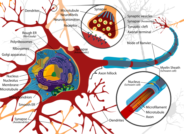
The brain, spine, and peripheral nerves are made up mostly of specialized cells called neurons and glia. Neurons are cells that sense their environment and send signals as a result. In combination, they are capable of creating all that we know as reality, including our pain. There is a wide variety of neurons in the body serving different functions, but we can use Figure 1: A neuron1 as a general illustration. (It is also an example of an attractive technical drawing). The bodies (or somas) of neurons are highly variable in size. They range from 4 to 100 micrometers (millionths of a meter) in diameter. If we assume an average 25 micrometers as a size, about 130,000 could be laid out in a single layer on a square inch, and 20,000,000 cell bodies would fit into a cubic inch.
Neurons are highly variable in a number of other ways. A basic form for a neuron is shown in figure 1. Neurons specialize in generating and sending signals. A dendrite (the input to the neuron) can be sensitive to any of a number of conditions: physical pressure, the presence of certain chemicals, light, or signals from other neurons. Stimulation of the dendrites causes the neuron to send a signal down its axon to synapses at the end(s) of the axon. At the end of the axon are usually multiple endings that are designed to pass signals on to other cells. These endings are specialized structures called synapses. They work by releasing tiny amounts of specific chemicals, called neurotransmitters, into the area between the sending neuron's axon and sensitive areas of the cell that receives the signal. A synapse is shown in one of the insets in Figure 1.
The receiving cell contains tiny chemical structures on its surface called neuroreceptors that are designed to respond to specific types of neurotransmitters. When enough neuroreceptors receive enough released transmitter molecules, it will cause the receiving cell in turn to take action. If the receiving cell is another neuron, the receiving neuron will fire its own signal. In this way, neural signals can propagate through the nervous system, allowing the nervous system to do its work, which in many ways is similar to the work of an electronic computer. (More details about how complex behavior can result from these simple components are described in a following section, Neural Circuits.)
The receiving cell may not be another neuron, but may instead be a muscle cell, an endocrine gland, or a variety of other cell types. In these cases the receiving cell will take an action suited to its type. A muscle may contract or an endocrine gland may release chemicals into the bloodstream.
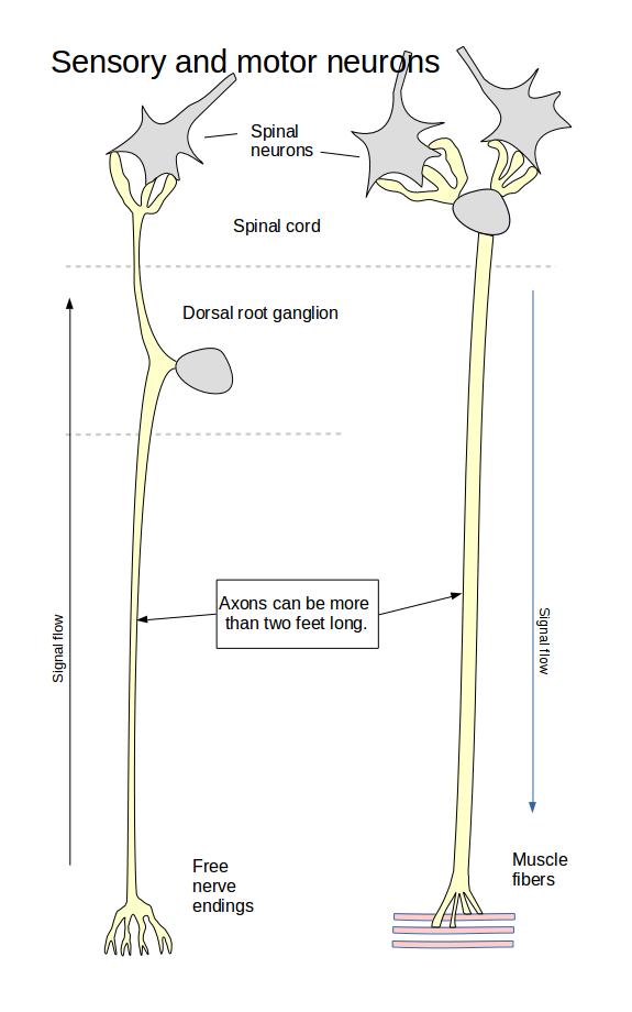
Since neurons perform a range of different functions, they also differ in their physical form. Figure 2, for example, shows schematically the shape or morphology of a sensory and a motor neuron. On the left in the diagram is a sensory neuron, the type of neuron that carries sensory input from, for example, the skin to the spine. The axon of such a neuron can be quite amazingly long. The sensory nerves that serve the big toe, for example, enter the spine at about the level of your waist, so they may be more than two feet long. The axon, the long part of the neuron, isn't built up from multiple cells. It is a part of a single cell.
A sensory neuron extends from within the spinal cord to some part of the body. The sensory neuron in Figure 2 is shown ending with free nerve endings, which generate most normal pain from the body when they are stimulated by pressure, chemicals, or certain other conditions. The axon, shown in yellow, extends toward the spinal cord and passes its own cell body or soma, which is shown here in grey. The cell bodies of all the sensory neurons are contained in groups called dorsal root ganglia. The axon continues into the spinal cord where it branches into dendrites that form synapses with neurons in the spinal cord. Thus the free nerve endings signal the spinal neurons when they are excited.
On the right in the same diagram is a motor neuron. A motor neuron is a neuron whose role is to trigger actions in other non-neural cells, including muscles. It is similar to the sensory neuron in its general shape. While the sensory neuron is meant to carry signals into the spine, the motor neuron's job is to carry signals out of the spine to its target muscle or other target cells.
Unlike sensory neurons, the cell bodies of motor neurons are inside of the spinal cord. Spinal neurons, shown in gray, form synapses with the dendrites that emerge from the motor neuron's cell body. These dendrites are shown in yellow. The body of a motor neuron transfers signals into the cell's axon. The signals continue through the axon, which branches into a number of dendrites. These form synapses with, for example, muscle fibers. The signals that originated in the spine cause the muscle fibers to contract.
While the diagram shows a sensory nerve with eight endings, typically they have from dozens to hundreds of endings. Similarly, the motor neuron shows four endings attached to two muscle fibers. Motor neurons have connections to from ten to thousands of muscle fibers, depending upon the muscle.2
Figure 2 shows two forms of central nervous system neurons. There is a great variety of neuron shapes within the central nervous system. Modern neuroscience methods have revealed that our CNS is not made of homogeneous general-purpose "intellegent" tissue, but instead it is highly structured into what can be called modules, each with specific computational roles to perform. (More about this in a later section, The Brain's Architecture and its Role in Pain.) Form follows function, so short, highly-branched axons are found where high connectivity is needed, while long axons are found where communication over long distances is needed.
The signal carried by a neuron is called an action potential. The name refers to the electrical nature of the signal. (An electrical “potential” is measured in volts). Although an action potential does have an electrical aspect, it isn't the same as an electric current running through a wire. An action potential uses the movement of ions through the cell membrane of the neuron to propel itself along the neuron's dendrites and axon, while a wire uses the movement of electrons. For that reason, an action potential moves much more slowly than a signal through a wire. Nevertheless, the action potential generates voltages that can be read by an electrocardiogram (which reads heart rhythms), an electromyelogram (which reads action potentials moving through muscle), or an electroencephalogram (which reads action potential moving through the outer areas of the brain).
Neurons contain ions in their internal fluid substance, which is called cytoplasm. An ion is a molecule that has an electric charge. Neurons are also surrounded by the extracellular fluid that bathes all the body's cells. The extracellular fluid also contains ions. Both cytoplasm and extracellular fluid are mostly water. The ions that are most important in action potentials are small ions that move easily and quickly in the intracellular and extracellular fluids and that carry a large electrical charge for their size and weight. These ions include sodium, chlorine, potassium, and calcium. Neurons transmit signals along their length by moving ions into and out of the cell along the length of the cell membrane. Where an ion moves, so moves its electrical charge, so that the neural signal can be sensed by electronic devices.
When a nerve signal passes across a synapse from one neuron to another, however, it is passed by the release and sensing of neurotransmitters. Both of these processes takes a certain length of time and requires that the cell expend energy. Because of this, neural signals travel much more slowly than an electrical signal through a wire does, which moves at nearly the speed of light. In addition, neurons generally require time to prepare to send their next signal. These two factors mean that 1) there is a delay between the occurrence of an event and its being sensed in the CNS; 2) there is a limit to how much information can be carried over a neural transmission route. (This is explained in more detail in From Nociceptors to the Brain.) Neural signaling is more like an old-fashioned telegraph message than modern digital computing technology. (Technically, you can say that neural signaling has limited bandwidth and high latency.)
The speed of neural transmission depends on the types of neurons that carry the signals. Just as in 21st century technology, speed costs resources, so your nervous system has multiple channels that carry sensory information to the brain at different speeds. This requires the CNS to compensate for the arrival at different times of information relating to the same sensed event. These and other characteristics of pain physiology provide the opportunity for problematic behavior of the pain system. These characteristics and behaviors are further described in Pain Science 2: Nociceptors and the Spine.
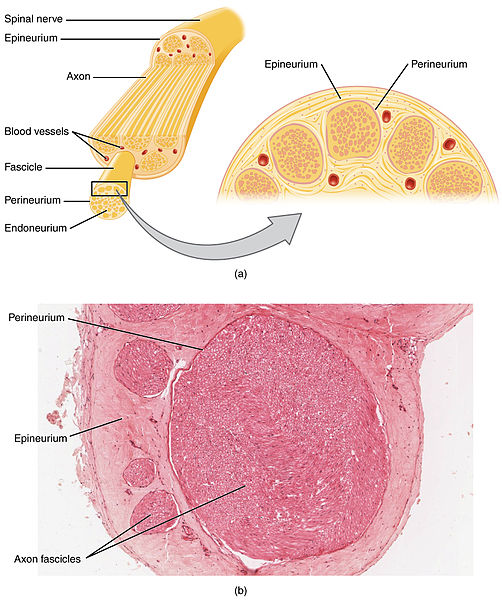
So far I've been discussing neurons, individual nerve cells. They are complex, varied, and specialized. A nerve, on the other hand. is an organ of the body outside of the central nervous system that contains and protects usually multiple neural axons. Figure 3 shows a diagram and a photomicrograph of a nerve. Large nerves, such as the spinal nerves, contain the axons of thousands or tens of thousands of motor and sensory neurons. The neural cell bodies for these nerves are within the spine (for motor nerves), or in dorsal root ganglia just outside the spine (for sensory somatic nerves).
The axons within a nerve are protected by layers of connective tissue (the endoneurium, perineurium, and epineurium in the diagram) and are grouped according to their destination within the body. Large nerves branch repeatedly until finally individual axons emerge.