Last updated: Mon, Jan 20, 2025
…we emphasize the inherent complexity of the spinal and supraspinal interconnections that must be involved in the human experience of pain, which remain to be fully elucidated.1
This section describes the parts of the brain that are known to be involved in pain processing, some of their connections, and their functions.
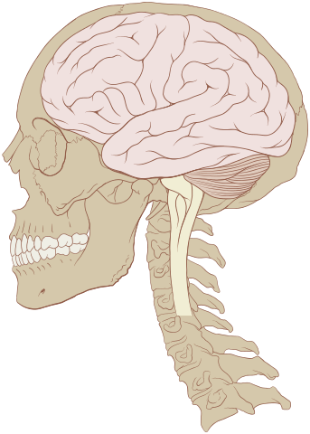
The brain occupies most of the skull above and behind the eyes and mouth. It is estimated to contain 80-90 billion neurons (nerve cells). Its most conspicuous feature when seen from the outside is the cortex (or cerebral cortex). See Figure 1: The human brain2. The cortex is a heavily folded sheet made up of regular layers of neurons. The cortex is sometimes referred to as “gray matter” because (so I have read) it appears gray, since it is made up largely of the cell bodies of neurons. Areas beneath the surface are “white matter” because they are made up largely of the fibers that connect neurons, which are covered with a fatty white protein, myelin.
There are many ways to break down the brain into a hierarchy of sections. One of these is based on the embryologic development of the brain. Since that perspective is not essential to understand the processing of pain, I'll use a simpler breakdown that is based not on embryology or evolutionary development, but on location and function.
Folded within the cortex, on the bottom side of the brain cavity, are the thalamus and the hypothalamus. The thalamus is a communications hub of the nervous system. Information from most of the somatic sensory systems, excluding the olfactory (smell) system, passes through it to the cortex. The thalamus is also know to be involved in wakefulness and attention, and is connected to the hippocampus, which is involved in memory. The hypothalamus is another communications hub, but processes sensory information from the viscera. The hypothalamus is also centrally involved in affect or emotion. The thalamus and hypothalamus together make up the diencephalon.
The diencephalon is surrounded by a series of structures that are known as the limbic system. The limbic system is involved in emotion, motivation, and memory.
Behind or below the diencephalon (lower in the brain) is the brain stem. Although the brain stem appears to be an extension of the spinal cord, it is usually viewed as part of the brain. The brain stem is involved in controlling autonomic functions (see Pain Science 4: Partner Systems) and in processing and relaying signals to and from the body.
The areas of the brain are highly interconnected, and although much of it appears to be undifferentiated, it is in fact made up of areas with specialized functions and connected in a way that is consistent from individual to individual. Today's scientists have a number of techniques that allow them to learn about the connectivity and functions of different areas of the brain. These techniques are described in Studying the Brain.
Notwithstanding the "inherent complexity" of the brain mentioned in the starting quotation, there are some generalizations about brain structures and their functions that should be kept in mind.
While there are numerous tracts of neural axons that connect distant parts of the brain, brain neurons are mostly connected locally, and connect to a small number of neurons. Because of this, what particular neurons do depends on the local assembly of neurons that they belong to. What assemblies of neurons do depend on what other assemblies they are connected with through neural tracts. The function of a functional group of assemblies depends on its component assemblies.3 These principles are the basis for the idea of functional structures in the brain.
Similarly, the brain for the most part isn't made up of localized "centers" that perform final functions, but rather of systems composed of interconnected assemblies. Thus, for example, there is no "pain center" in the brain.
The brain tends to process "raw input" from each of the sensory systems in separate specialized systems. For example, a large area of the cortex at the back of the skull is dedicated to making sense out of visual input. It includes specialized structures for uses such as detecting edges. While there are areas for integrating edge data with other visual comclusions to form integrated images, and there areas for filtering and combining auditory information into images of speech, there is no general association area of the brain that is able to integrate all forms of data that the brain can create.4
So it is with pain. Multiple brain areas are involved in creating the experience of pain, and the pain experience is not a simple sensation or a simple emotion, but a phenomenon with a number of dimensions.
The somaesthetic system is a unitary, integrated system comprised of specialized component parts. Several parallel systems analyse the input simultaneously to bring about the richness and complexity of pain experience and response. Some areas are specialized to select sensory-discriminative information while others play specialized roles in the motivational-affective dimension of pain. These parallel information-processing systems interact with each other, and must also interact with cortical activities which underlie past experience, attention, and other cognitive determinants of pain. These interacting processes produce the myriad patterns of activity that subserve the varieties of pain experience.5
Within this section...
The Ascending Tracts (Last updated: Wed, Feb 19, 2025)
The Brain Stem (Last updated: Tue, Mar 4, 2025)
The Pain Matrix (Last updated: Tue, Feb 18, 2025)
The Descending Tracts and Descending Pain Modulation (Last updated: Fri, Mar 7, 2025)
Neurotransmitters Involved in Pain Processing (Last updated: Tue, Mar 4, 2025)
The Pain Matrix in Chronic Pain States (This page is incomplete.)
Or skip to...
Pain Phenomena Involving the Brain (Last updated: Sun, Mar 9, 2025)