Please use the form below to submit comments. Also provide an e-mail address and name. Your e-mail address and/or name will be used only to communicate with you about this or future comments you may submit. I am particularly keen to receive references to published material that contradicts the assertions and arguments I have made.
By submitting the above comment, I grant to Ross Alan Hangartner the right to incorporate the comment in full or in part, literally, paraphrased, or conceptually, as he sees fit, into State of Pain or other writings that he may create in the future. However, I don't grant permission to include my name or e-mail address, or to use them in any other way than to contact me for follow-up. I understand that by submitting the comment I acquire no right of any kind in State of Pain or other writings of Ross Alan Hangartner.
Last updated: Fri, Mar 21, 2025
So far in this work I've mostly been describing the somatic nervous system, which controls the voluntary movements of the body and detects/creates pain of the musculoskeletal system, among other functions. Your autonomic nervous system (ANS) is a second division of your nervous system. If "autonomic" reminds you of "automatic," it should. A distinguishing feature of the ANS is that its function is largely automatic. It works without your conscious awareness. On the other hand, you are often aware of its effects, which you sense as interoception.
The ANS shares the brain, the brain stem, and to some extent the spine, with your somatic nervous system. Outside of the spine, the ANS largely has its own nerves and its own functions, but shares spinal nerves with the somatic nervous system. It serves the general function of regulating the level of arousal of your body. The ANS has both sensory (afferent) neurons, and motor (efferent) neurons. In the somatic nervous system, motor neurons activate voluntary muscle fibers--the skeletal muscles. In the ANS, motor nerves activate involuntary or smooth muscles, such as those that control the tightness of blood vessels, or cause viscera to perform certain actions.
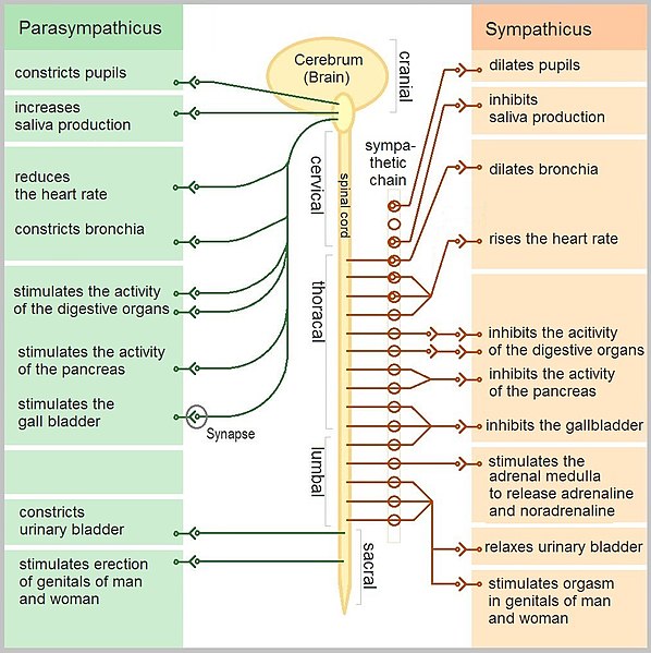
The diagram is an abstract representation of the ANS. In the center panel of the figure is a diagram of the brain and spinal cord. You can see a number of dark nerves leaving the brain stem and the sacral spinal cord. These connect to the left panel of the figure. These represent parasympathetic nerves. In the left panel, some of the actions of the parasympathetic division of the ANS or parasympathetic nervous system are listed.
In the center panel again, but on the right side, you can see a number of red nerves leaving the spine at thoracic and lumbar levels. These represent sympathetic nerves. In the right panel some of the actions of the sympathetic nervous system are listed.
If you compare the parasympathetic functions to the sympathetic functions, you'll see that the parasympathetic functions tend to be appropriate for times of low stress or threat, while the sympathetic functions tend to be appropriate for times of stress or threat. The early conception of the sympathetic and parasympathetic branches was based on this distinction, but that has been found to be an oversimplification.
There are twelve pairs of nerves that leave the brain and brain stem and pass into the body. These are the cranial nerves. Four of the twelve carry parasympathetic neurons, those designated as III, VII, IX, and X. Cranial nerve X, the vagus nerve, is particularly important. The vagus contains parasympathetic motor nerves to the organs from the neck down to the transverse colon. It is responsible then for heart rate, digestive system peristalsis, sweating and control of bodily inflammation through its connection to the spleen.
The red sympathetic nerves on the right side of the center panel exit the spine through the dorsal root ganglia, then separate from somatic nerves and connect to form the sympathetic chain, which runs outside and along the spine, and includes a number of ganglia in which spinal sympathetic neurons connect with extraspinal, postganglionic sympathetic neurons which in turn connect to organs that are affected by sympathetic input. The sympathetic neurons that emerge from the different segments of the spinal cord are not distributed to the same part of the body as the somatic spinal nerve fibers from the same segments. While the sympathetic chain appears on the right side in the diagram, in the body it is bilateral.
Some of the postganglionic motor fibers pass back from the sympathetic chain and rejoin spinal nerves at all levels of the cord. These sympathetic fibers are all very small type C fibers. They extend through the spinal nerves to all parts of the body. These control the blood vessels, sweat glands, and piloerector muscles of the hairs. About 8 per cent of the fibers in the average spinal nerve are sympathetic fibers.1
Your autonomic nervous system is also involved in pain. It can generate pain on its own, and it can affect pain that originates in the somatic nervous system. The ANS is heavily involved in creating or influencing many of the "co-morbidities" associated with pain, including stress. (Co-morbidities.) When the ANS is exposed to extended painful conditions, particularly inflammation, the ANS can provoke an illness response that subdues your entire mood and can last as long as the painful conditions.
Within this section...
Physiologic Functions of the ANS (Last updated: Sat, Mar 15, 2025)
ANS Pain Circuits (This page is incomplete.)
Autonomic Pain Phenomena (This page is incomplete.)
Or skip to...
Pain and the Sickness Response (This page is incomplete.)