Please use the form below to submit comments. Also provide an e-mail address and name. Your e-mail address and/or name will be used only to communicate with you about this or future comments you may submit. I am particularly keen to receive references to published material that contradicts the assertions and arguments I have made.
By submitting the above comment, I grant to Ross Alan Hangartner the right to incorporate the comment in full or in part, literally, paraphrased, or conceptually, as he sees fit, into State of Pain or other writings that he may create in the future. However, I don't grant permission to include my name or e-mail address, or to use them in any other way than to contact me for follow-up. I understand that by submitting the comment I acquire no right of any kind in State of Pain or other writings of Ross Alan Hangartner.
Last updated: Fri, Jul 26, 2024
The spinal cord is a cylindrical structure made up of neurons and cells that support them. It joins the base of the brain stem after it leaves the skull and extends down the spine. The spinal cord is protected by the bony arches that make up the dorsal (back) portion of each of the spinal vertebral bones. The cord itself extends to just below the bottom ribs. Individual spinal nerves continue beyond that point. The membranes that enclose the spinal cord extend down into the sacrum.
The spinal cord is the main conduit for communication between the body and the brain. Although the spinal cord and the brain stem are considered to be separate organs, the brain stem sends fibers into the spinal cord and vice versa. Signals that travel toward the brain are called afferent signals, and the neurons that carry them are called afferent neurons. Afferent signals include all of the sensory signals from the body. Signals that travel away from the brain are called efferent signals, and the neurons that carry them are efferent neurons or effectors. Examples of efferent signals are signals to muscles to contract or to the heart to tell it to pump harder. Not all of the efferent signals go to muscles or internal organs--some signals from the brain and brain stem go to neurons within the spine, as we will explore later (Ascending Projection Neurons and Descending Projection Neurons).
Twenty-six pairs of spinal nerves emerge from the spinal cord. Each pair of spinal nerves innervates a defined strip of skin and a defined set of muscles and other deep tissue. This is one instance of a general principle in the organization of the nervous system: Nerves and nerve fibers are arranged to reflect the arrangement of the structures that they innervate. This is called somatotopic organization. (“Somatotopic” means “place in the body”.)
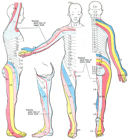
A dermatome is a portion of the outer surface of the body that is innervated by a particular pair of spinal nerves. Although diagrams such as Figure 1: Dermatomes1 show the dermatomes as distinct bands of skin, the boundaries are in fact fuzzy and overlapping. Nevertheless, doctors can use the dermatomes to localize nerve problems to a specific spinal nerve.
A myotome is a group of muscles that are innervated by a single spinal nerve. A muscle may be innervated by more than one spinal nerve. Myotomes unfortunately don't make clear pictures. Example myotomes include:
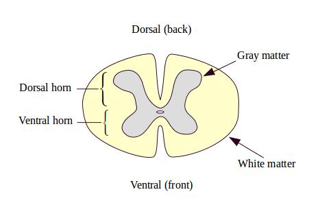
Your spinal cord varies in width from about the size of your little finger to about the size of your thumb. It is organized into segments, and has a similar appearance and organization at each level. Figure 2: Cross section of the spinal cord is a generic cross-section of the cord. It is composed of white matter on the outside with a central area of gray matter. The white matter consists largely of myelinated axons. These axons carry signals up and down the cord, between the cord and the brain, and between different levels of the cord. This includes sensory information, which primarily goes up toward the brain, muscle control signals, which primarily go down the cord toward the periphery, and autonomic nervous system signals, which go in both directions.
The gray matter of the spinal cord is gray because it contains a lot of neuron bodies. Each segment contains several million neurons and also contains many times that number of small immune system cells called astrocytes and microglia. (See The Central Nervous System Environment) The gray matter is considered to have two major parts, the dorsal horn and the ventral horn. (They were given the name “horns” because they have a somewhat horn-like shape in cross section.)
The ventral (front) horn is concerned with processing efferent signals, those that travel from the central nervous system to the body. The bodies of motor neurons (the neurons that direct muscles to contract) are in the ventral horn. The axons of the motor neurons along with the other efferent neurons emerge from the ventral horns on either side of the cord, forming the two ventral roots of the spinal nerves at that level.
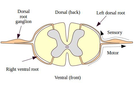
The dorsal (back) horn is concerned with processing afferent signals, that is, signals that travel from the body to the central nervous system. The soma (cell bodies) of these sensory neurons all live outside the spinal cord in a swelling called a dorsal root ganglion. The axon of a sensory neuron travels past its soma into the spine, where it forms synapses with neurons of the dorsal horn. Figure 3: Spinal nerve roots shows a cross section of the cord with the spinal nerve roots.
The organization of interneurons in the dorsal horn is very complex, and we still know little about the neuronal circuits in which they take part.2
The somatic sensory neurons form their first synapses in the dorsal horn, so this is where the initial processing of signals that result in pain occurs. Four types of neuronal components have been found in the dorsal horn: