Please use the form below to submit comments. Also provide an e-mail address and name. Your e-mail address and/or name will be used only to communicate with you about this or future comments you may submit. I am particularly keen to receive references to published material that contradicts the assertions and arguments I have made.
By submitting the above comment, I grant to Ross Alan Hangartner the right to incorporate the comment in full or in part, literally, paraphrased, or conceptually, as he sees fit, into State of Pain or other writings that he may create in the future. However, I don't grant permission to include my name or e-mail address, or to use them in any other way than to contact me for follow-up. I understand that by submitting the comment I acquire no right of any kind in State of Pain or other writings of Ross Alan Hangartner.
Last updated: Sun, Jun 18, 2017
There are many kinds of sensory neurons in your body. Sensory neurons have receptors in their terminal branches that respond to stimulation from its environment. The type of stimulation that a receptor responds to (pressure, temperature, chemicals) is called the receptor's modality. Table 11 contains a list of receptor types.
Since we are interested in pain, we are interested in a subset of the sensor types listed in Table 1, those that innervate the body and are called somatosensory neurons.
Mechanoreceptors (respond to touch or pressure)
|
Thermoreceptors (respond to heat or cold)
|
Electromagnetic receptors (respond to light)
|
Chemoreceptors (respond to chemicals)
|
The receptor types that are most important for pain are the mechanoreceptors, thermoreceptors, and chemoreceptors. There is no separate category of “pain receptors.” The closest we will find to a pain receptor is a nociceptor, meaning a receptor that responds most strongly to noxious stimuli, that is, extreme pressure or temperature, or certain chemicals associated with tissue damage and inflammation.
All of these receptor types ‘fire’ by raising the excitation level of the neuron, which they do by admitting positively-charged ions into the neuron. The mechanoreceptors do this when they are physically distorted by forces in the tissue. The mechanoreceptors are ion channels that open when they are stretched. Thermoreceptors become more permeable when the temperature changes from normal body temperature. Chemoreceptors open in the presence of particular chemical compounds.
All of the sensory neurons signal their activated condition by firing action potentials. In general, the more intense the sensed condition, the more frequently an action potential is fired.
There are several reasons for the large number of subtypes within the class of mechanoreceptors. Some of the types are highly specialized for a purpose, such as the muscle spindle receptors that monitor the length of muscles or Golgi tendon receptors that monitor the tension in tendons. Among the skin sensors, some have specialized endings that allow detection of particular conditions of the skin or flesh. A Pacinian capsule, for example, is built so that it produces a signal while it changes shape, but stops signaling almost immediately when the deformation stops. In this way, it signals vibration and pressure. The Meissner corpuscle is a slightly different configuration that is sensitive to a lower frequency of vibration. Merkel receptors are made up of a special Merkel cell in conjunction with a terminal nerve ending, and produce repeated signals in response to sustained pressure.
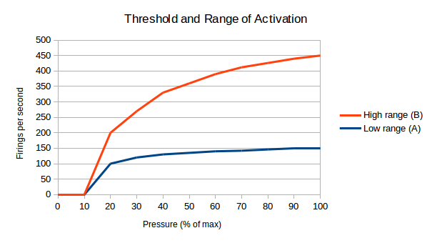
Two important parameters of the mechanoreceptors are their activation threshold and their adaptation characteristics. The activation threshold is the minimum pressure required to generate an action potential. Mechanoreceptors can be loosely categorized into low-threshold and high-threshold receptors. Receptors with the same activation threshold can have different response characteristics. Figure 1: Mechanoreceptors with different ranges of activation illustrates this idea. Both receptor A (in blue) and receptor B (in red) are activated at a low threshold, 10% of their maximum. Receptor A reaches its maximum firing rate at a fairly low pressure, while the signal of receptor B varies between a very low and a very high pressure.
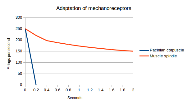
Adaptation refers to the way a sensor's signal varies when it’s stimulated continuously. A sensor whose output in response to a continuous stimulus falls off rapidly is said to “adapt rapidly.” Figure 2: Adaptation of mechanoreceptors2 illustrates this idea, comparing the output of a Pacinian receptor with the output of a muscle spindle receptor. The Pacinian receptor falls silent very soon after it is deformed, while the muscle spindle sensor continues to fire action potentials for as long as the muscle is stretched. The Pacinian receptor therefore reports that a change has happened, while the muscle spindle receptor reports that something is continuing. Many of your nociceptors adapt slowly or not at all--they continue firing until the condition that trggered them changes.
Besides differences in modality (pressure, temperature, chemicals), threshold, and sensitivity, sensory neurons differ in their chemical makeup. Certain types of sensory neurons release different transmitters depending on what type of tissue they innervate.
Within this section...
Three Principal Types of Sensory Neurons (Last updated: Sun, Jun 18, 2017)
Or skip to...
Anatomy and Distribution of Sensory Nerves (This page is incomplete.)