Please use the form below to submit comments. Also provide an e-mail address and name. Your e-mail address and/or name will be used only to communicate with you about this or future comments you may submit. I am particularly keen to receive references to published material that contradicts the assertions and arguments I have made.
By submitting the above comment, I grant to Ross Alan Hangartner the right to incorporate the comment in full or in part, literally, paraphrased, or conceptually, as he sees fit, into State of Pain or other writings that he may create in the future. However, I don't grant permission to include my name or e-mail address, or to use them in any other way than to contact me for follow-up. I understand that by submitting the comment I acquire no right of any kind in State of Pain or other writings of Ross Alan Hangartner.
Last updated: Tue, Nov 19, 2024
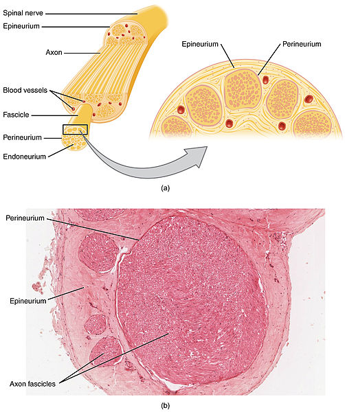
Each of the spinal nerves is enclosed in a membrane that separates it from the surrounding tissue. The nerves branch repeatedly until individual neurons emerge. Each spinal nerve contains three functional types of axons: 1) motor axons that cause muscles to contract; 2) sensory axons; and 3) sympathetic axons that control functions such as the size of arteries and sweating. See Figure 1: A spinal nerve1 for the structure of a large nerve.
Each sensory axon travels the entire distance from the spinal cord to the neuron's sensory receptors. The tiny axon of each of the tens of thousands of nerves that go to the foot can be almost a meter long.
The cell bodies of all of the sensory neurons in a spinal nerve are contained in a single node, nestled between the vertebrae where the nerve emerges from the spinal cord, called the dorsal root ganglion.