Please use the form below to submit comments. Also provide an e-mail address and name. Your e-mail address and/or name will be used only to communicate with you about this or future comments you may submit. I am particularly keen to receive references to published material that contradicts the assertions and arguments I have made.
By submitting the above comment, I grant to Ross Alan Hangartner the right to incorporate the comment in full or in part, literally, paraphrased, or conceptually, as he sees fit, into State of Pain or other writings that he may create in the future. However, I don't grant permission to include my name or e-mail address, or to use them in any other way than to contact me for follow-up. I understand that by submitting the comment I acquire no right of any kind in State of Pain or other writings of Ross Alan Hangartner.
Last updated: Sun, Jun 18, 2017
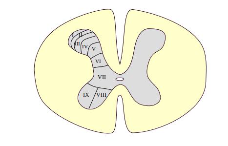
The organization of the spinal cord has had much attention from researchers, and has been a difficult puzzle to solve. One of the early clues came when a researcher noticed that the horns of the spinal gray matter form layers. These layers are formally called the laminae of Rexed after their discoverer. Some of the layers are visible to the naked eye. Most are apparent only under an optical microscope with the aid of tissue staining and other histological and pharmacological techniques. Figure 1: The laminae of Rexed shows nine laminae in the left horn.
The different layers have specialized functions.