Last updated: Tue, Feb 11, 2025
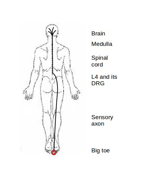
The diagram shows schematically how pain-producing neural signals travel through the nervous system. If you should stub your big toe, receptors at the end of at least one sensory neuron will send signals up the axon, which will join the spinal nerve L4. When the signal nears the spine, the signal passes the cell body, which is in the L4 dorsal root ganglion.
The axon continues into the spinal cord, where it forms synapses with CNS neurons inside the spine. There, the spinal neurons process the toe signal, and, assuming that the signal results in pain, the signal passes to the long neurons that ascend the spine and into the brain.
The spinal cord stops and the medulla oblongota or brain stem begins just below the skull. The cord and the medulla appear continuous to the naked eye, but they perform different processing functions on the toe signal. The signal then passes through and spreads out to a number of structures of the brain, including the midbrain, the limbic system, the cerebellum, and the cortex. These major components of the brain and the numerous smaller structures of which they are composed each have different functions in interpreting and acting upon the signal that originated in your toe.
The view of the pain system that is shown in Figure 1 is simplified in at least two ways. First, it shows only a signal from a single sensory neuron. In reality, the spinal cord and the brain are continually receiving millions of signals from hundreds of type of neurons that report on conditions from virtually all the tissues of the body. The spine and brain must make sense of all these signals and initiate actions to respond appropriately. Most of the input to the CNS does not enter consciousness.
Second, the response of the CNS to its inputs results not only in your conscious awareness of situations such as a stubbed toe, but in adjustments to the CNS itself. This last point is key to understanding the nature of pain and its effects on you.
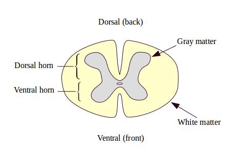
Figure 2 shows a cross-section of the spinal cord. There is one of these sections or segments for each of the spinal nerve pairs. (There are analagous structures in the brain stem for the cranial nerves that originate there.) Each segment is less than half an inch thick. The sensory nerves, such as the big toe nerve that signals your stubbed toe, enter through the dorsal root ganglion, and pass into the gray matter in the dorsal horn of the spinal cord. The neural tissue in the center of the spinal cord is grey because it contains mostly neural bodies and dendrites. The dorsal horn is built for connectivity. It routes signals within the spinal cord, and it routes signals to the brain. It also performs active processing on neural signals. (This is discussed in more detail in Pain Science 2: Nociceptors and the Spine.)
Once the incoming signal has been processed in the cord's grey matter, it will be relayed up the spine through the spinal white matter. (Shown here in yellow.) White nervous tissue is pale because it contains largely neural axons which can be encased in myelin, a white waxy substance that is present on axons that communicate over considerable distances. The spinal cord's white matter is made up mostly of fast-conducting myelinated axons.
In contrast to sensory neurons, the cell bodies of motor neurons, which carry signals out of the central nervous system, are contained within the spinal cord, in the grey ventral horns. Cell bodies of the autonomic nervous system are found in the lateral horns of the spinal grey matter. (Not shown in Figure 2.)
The spinal cord performs several kinds of processing of sensory signals. Several types of reflexes are triggered mostly within the spinal cord.
The central nervous system must track where in the body axons come from or go to. It does this by keeping the endings of nerves from areas close together in the body close together in the spine and brain. This is called topographical organization. When sensory nerve endings first enter the dorsal horns they are topographically organized, and they are repeatedly organized that way wherever the CNS needs location information. Figure 3 represents the topographic organization of the neurons at one of the major structures in the brain's cortex. You may have seen this image before. The size of body parts are reduced or enlarged in this diagram to show that the brain contains higher spatial resolution for some parts of the body than for others. The topographically-organized nervous structures are sometimes called "homunculi" (singular, "homunculus").
These topographical maps or homunculi aren't fixed structures. They can be changed by how you use your body. If a particular body area is used a lot, its representation in the homunculus grows. If it is under-used, its representation shrinks. Such changes can cause the map to lose its correspondence to the body and the peripheral nerves, which can cause the pain system to make an incorrect interpretation of where sensory signals originated, or to send motor instructions to the wrong muscles or glands.

Another important processing function that the spinal cord participates in is regulation of the sensitivity of the sensory system. The nervous system regulates its sensory sensitivity much as it regulates body temperature, salt concentrations, or the light sensitivity of your retinas. The adjustability is an important factor in understanding the behavior of the pain system in both normal and abnormal operation.
The brain is a complex organ and its processing of sensory signals is complex. Pain is a product of the brain, not of the sensory neurons or of the spinal cord by themselves. So it's always true that pain is in your head. So also, in a very real sense, are your other feelings and sensations. When you feel a localized pain sensation, an emotional feeling accompanies it. So pain is characterized not only by an intensity and a location, but also by an aversive emotion and potentially by other feelings or emotions. Pain is not a simple sensation.
As Figure 1 shows, pain signals are sensed by multiple areas in the brain, along with all the other sensory input from your body. The nervous system is a parallel-processing system in which processing must be coordinated, both logically and in time, in order to make sense out of the many signals it is always receiving.
One of the results of the pain system is that your toe hurts when you stub it. But it only feels as if the pain is in your toe. Pain is the result of interactions between your central nervous system and the "raw" sensory input of your sensory nerves. The sensitivity of each component in the pain processing chain is constantly under adjustment.
Your pain system is complex and routinely creates illusions, such as the illusion that the pain itself is "in" the stimulated part of the body. Nevertheless, it is an orderly system. Understanding it in greater detail can help to understand why and how it sometimes seems to do more harm than good, how it is sometimes misleading, and, importantly, it sets limits to how it can be imagined to function. (See Pain Science 3: Neuroscience and the Brain for more.)